Our Services
Learn more about the services offered by Palm Beach Cardiology Center… Thanks!
Cardiac Event Monitoring
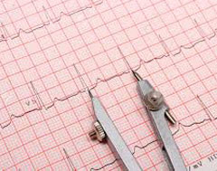
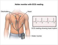
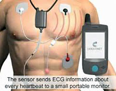
These devices provide real time, wireless data to a remote center and are used for diagnosis of arrhythmias that occur infrequently or for conditions that require prolonged monitoring. PBCC provides a full range of monitoring such including single lead patch devices for greater comfort and small implantable devices.
Echocardiography
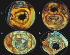
An echocardiogram uses ultrasound waves to assess the structure and function of the heart and its components, similar to a baby ultrasound. It uses doppler technology, similar to a police radar gun, to assess for valve integrity and hemodynamics. We are proud to offer expertise in 3 dimensional echocardiography and tissue tracking modalities that can be used for diagnosis and treatment of advanced heart conditions. We are dedicated to the highest quality standards in cardiac imaging and are proud to be one of the few centers in North Palm Beach that have an ICAEL accredited laboratory.
Vascular Ultrasound
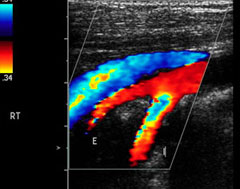
Vascular ultrasounds use ultrasound waves to detect blockages in blood flow that lead to decreased flow to certain body parts (brain, legs), can assess for weakening of the lining of blood vessel walls that can cause aneurysms. Sonography of the veins can be used to detect for blood clots or venous insufficiency that can lead to varicosities. We offer a full service of vascular sonography, including assessment of carotid, upper and lower extremity, and abdominal arteries, along with assessment of venous disease.
Stress Testing
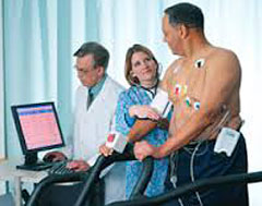
Cardiac stress test is a test used to measure the heart's ability to respond to external stress in a controlled clinical environment. They are most commonly used for assessment of the heart's own circulation (the coronary arteries) to meet the demands of the stress induced by exercise. The stress test can be performed with or without imaging for further assessment. An echocardiogram or a nuclear dye/camera can be used to assess the heart's blood flow demands in greater detail. For patients unable to exercise on a treadmill, we use a brief acting chemical agent to mimic the heart's response to stress.
Cardiac Catheterization & Angioplasty/Stents
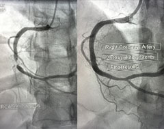
Cardiac Catheterization is a specialized study of the heart during which a catheter, a thin flexible tube, is inserted into the artery of the groin or arm. Under x-ray visualization, the tip of the catheter is guided to the heart. Pressures are measured and an x-ray Angiogram (Angio) movie of the heart and blood vessels are obtained while injecting a colorless dye through the catheter. X-ray movie pictures taken during the injection of the contrast material allow the coronary arteries to be visualized. If a severely blocked artery is identified that limits blood flow to the heart, this can be fixed with deployment of a balloon or stent. Stents are small expandable tubes used to treat narrowed or weakened arteries in the body. This otherwise known as a percutaneous coronary intervention or PCI. Direct visualization of the artery through ultrasound (IVUS) or pressure monitoring (FFR) can also be used to further assess the severity of disease and to establish procedural success.
Peripheral Arterial Interventions and Endovascular therapy
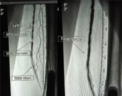
Peripheral artery disease is a common circulatory problem in which narrowed arteries reduce blood flow to your limbs.
When you develop peripheral artery disease (PAD), your extremities, usually your legs, don't receive enough blood flow to keep up with demand. This causes symptoms, most notably leg pain when walking (intermittent claudication).
Peripheral artery disease is also likely to be a sign of a more widespread accumulation of fatty deposits in your arteries (atherosclerosis). This condition may be reducing blood flow to your heart and brain, as well as your legs.
Venous Insufficiency and RFA
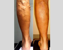
Healthy leg veins contain valves that open and close to assist the return of blood back to the heart. Venous reflux disease develops when the valves malfunction and no longer keep blood flowing in one direction out of the legs and back to the heart. This back flow of blood causes increased pressure on the leg veins. As a result, vein valves will not close properly, leading to signs and symptoms of varicose veins.
We offer a venous closure procedure that can be safely performed on an outpatient basis. Using ultrasound, we position the ClosureFAST™ catheter into the diseased vein through a small opening in the skin. The tiny catheter powered by radiofrequency energy delivers heat to the vein wall. As the thermal energy is delivered, the vein wall shrinks and the vein is sealed closed. Once the diseased vein is closed, blood is re-routed to other healthy veins.Following the procedure, a simple bandage is placed over the insertion site, and additional compression provided to aid healing.
Pacemaker/Defibrillators
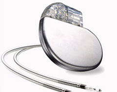
A pacemaker is implanted to treat bradycardia (an abnormally slow heart rate). Pacemakers can also adjust the heart rate to meet the body's needs, whether during exercise or rest. Implantation of a pacemaker involves positioning leads (thin, insulated wires) in the heart and placing the device in a pocket of skin, usually in the shoulder area. Typically the implant procedure involves only local anesthetics and a sedative, rather than general anesthesia.
An ICD (implantable cardioverter defibrillator), or defibrillator, helps stop dangerously fast heart rhythms in the ventricles (the heart's lower chambers). The device is used to treat sudden cardiac arrest and to restore a normal heartbeat. Implantation of a defibrillator involves positioning leads (thin, insulated wires) in the heart and placing the device beneath the skin, usually in the shoulder area. Typically the implant procedure involves only local anesthetics and a sedative, rather than general anesthesia. Most people have a fairly quick recovery after these procedures.

